How to Treat Subarachnoid Haemorrhage?
- January 12, 2024
- No Comments

What is Subarachnoid Haemorrhage?
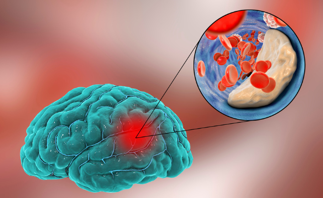
Subarachnoid hemorrhage (SAH) is a medical emergency marked by bleeding in the space between the arachnoid membrane and the pia mater enveloping the brain, known as the subarachnoid space. Typically caused by the rupture of an artery or vein in this region, it results in blood release into the cerebrospinal fluid. SAH demands immediate attention due to its potential severity.
This critical condition arises within the protective layers of the brain, encompassed by the dura mater, arachnoid, and pia mater. SAH is frequently triggered by head trauma or the rupture of a brain aneurysm, varying in severity levels. A key indicator is the sudden onset of a thunderclap headache, described as the most severe headache of one's life, distinguishing it from previous headache experiences.
Why is Subarachnoid Haemorrhage a Concern?
- Subarachnoid hemorrhage is a critical condition that can have severe consequences. The sudden and rapid accumulation of blood in the subarachnoid space can cause increased pressure on the brain, leading to a range of neurological symptoms. The most common cause of SAH is the rupture of a cerebral aneurysm, a weakened area in the wall of a blood vessel in the brain. Other causes may include head trauma, arteriovenous malformations, or certain medical conditions that affect blood vessels.
- The urgency in addressing subarachnoid hemorrhage is crucial because the increased pressure and damage to brain tissue can result in long-term neurological deficits or even be fatal if not promptly treated.
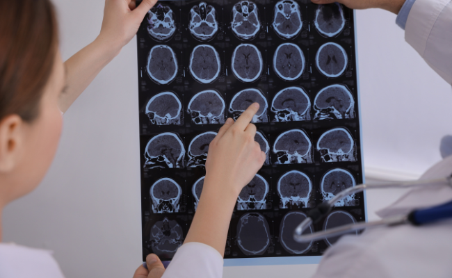
How to Diagnose Subarachnoid Haemorrhage?
- The diagnosis of subarachnoid hemorrhage typically begins with a thorough medical history and physical examination. However, the gold standard for confirming the diagnosis is usually imaging studies, such as a computed tomography (CT) scan or a lumbar puncture (also known as a spinal tap).
- A CT scan is often the initial imaging study performed, as it can quickly identify the presence of blood in the subarachnoid space. If the CT scan is negative, a lumbar puncture may be performed to analyze the cerebrospinal fluid for the presence of blood or other abnormalities.
- In some cases, additional imaging studies, such as magnetic resonance imaging (MRI) or cerebral angiography, may be ordered to further assess the underlying cause of the subarachnoid hemorrhage, such as the presence of an aneurysm.

Treatment Solutions for Subarachnoid Haemorrhage
Emergency Measures:
- Immediate stabilization is essential, and patients are often admitted to the intensive care unit for close monitoring.
- Blood pressure control is crucial to prevent re-bleeding, and medications may be administered to achieve this.
- Analgesics are often given to manage severe headaches associated with the condition.
Aneurysm Repair:
- If the subarachnoid hemorrhage is caused by the rupture of a cerebral aneurysm, prompt intervention is necessary.
- Surgical clipping and endovascular coiling are two common methods to repair an aneurysm.
- Surgical clipping involves placing a small metal clip around the neck of the aneurysm to prevent further bleeding.
- Endovascular coiling is a less invasive procedure where a catheter is threaded through the blood vessels to the site of the aneurysm, and coils are placed to seal off the aneurysm.
Vasospasm Prevention:
- Vasospasm, a potentially dangerous complication, can occur in the days following a subarachnoid hemorrhage.
- Medications like nimodipine are often administered to prevent vasospasm and improve blood flow to the brain.
Supportive Care:
- Patients may require various supportive measures, including respiratory support and intracranial pressure monitoring.
- Physical and occupational therapy may be initiated to help patients regain functional abilities.
Benefits of Timely Treatment for Subarachnoid Haemorrhage
Improved Survival Rates:
- Timely diagnosis and intervention significantly improve the chances of survival.
- Prompt treatment of the underlying cause, such as repairing an aneurysm, can prevent further bleeding and complications.
Reduced Neurological Deficits:
- Early intervention can help minimize the extent of brain damage, reducing the risk of long-term neurological deficits.
- Preventing vasospasm and ensuring proper blood flow to the brain contribute to better outcomes.
Prevention of Re-bleeding:
- Controlling blood pressure and repairing the source of bleeding, such as an aneurysm, are crucial in preventing re-bleeding.
- Swift action can mitigate the risk of recurrent hemorrhages and their associated complications.
Enhanced Quality of Life:
- Effective treatment and rehabilitation efforts can contribute to a better quality of life for survivors.
- Rehabilitation services play a crucial role in helping individuals regain lost functions and adapt to any remaining challenges.


Comments (0)
No comments yet